All About What Causes Bone Loss Around Dental Implants
The Main Reasons Bone Loss Happens Around Dental Implants
By Dr. Justin Wright, DDS
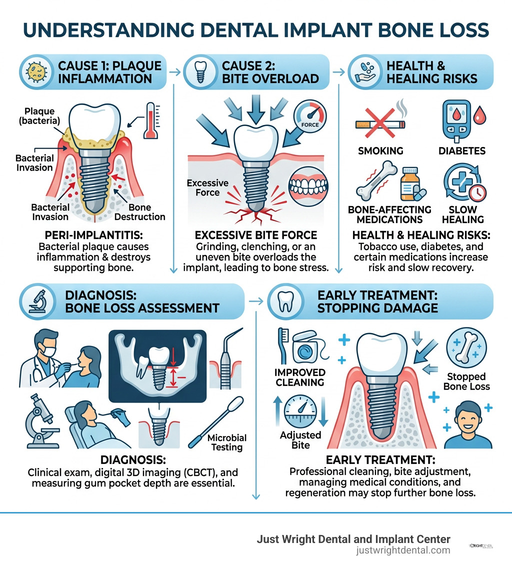
Bone loss around a dental implant is most often caused by peri-implantitis, an inflammatory infection driven by bacterial plaque around the implant. It can also result from too much bite pressure, poor implant position or restoration fit, smoking, uncontrolled diabetes, thin or limited starting bone, and problems with healing.
Some bone remodeling can occur as the jaw adapts after implant placement. Progressive bone loss is different. It needs an exam as soon as possible because early treatment may stop the damage before the implant becomes loose.
Key causes include:
Peri-implantitis: Plaque builds up below the gumline, causing inflammation that can destroy the bone supporting the implant.
Excessive force: Grinding, clenching, or an uneven bite can overload an implant. Unlike a natural tooth, an implant does not have a cushioning ligament.
Surgical or restoration issues: Implant depth, position, exposed threads, cement left beneath the gums, or a poorly shaped crown can make cleaning harder or place stress on the bone.
Health and healing factors: Tobacco use, poorly controlled diabetes, some bone-affecting medications, and low bone density can raise risk or slow healing.
Material-related inflammation: Titanium particles or corrosion may contribute to inflammation in some cases, especially when combined with bacterial plaque and mechanical stress.
I am Justin Wright, DDS, and over 18 years of placing more than 10,000 implants, I have treated many patients seeking answers about what causes bone loss around dental implants, including people with failed work completed elsewhere. The next sections explain how I look for the cause, measure the bone change on 3D imaging, and choose treatment based on what is actually happening around the implant.
What Causes Bone Loss Around Dental Implants?
Bone loss around dental implants occurs when the delicate balance between natural bone remodeling and destructive inflammatory stress is disrupted. In clinical practice, this breakdown stems from a combination of microbial plaque buildup, excessive physical biting pressure, poor surgical positioning, or an overactive immune response.
When an implant is placed into the jawbone, a biological process called osseointegration occurs, where living bone cells fuse directly to the titanium surface. In the initial healing phase, minor crestal bone remodeling often happens as tissues establish a biological seal. However, true marginal bone resorption happens when chronic irritation shifts the body into a state of continuous bone breakdown. Peer-reviewed research, including a scientific analysis of marginal bone remodeling, highlights that while initial remodeling settles near the first thread, ongoing loss indicates an underlying issue that requires clinical intervention.
To understand why bone recedes, we evaluate whether the trigger occurred during the early healing window or developed years later due to biological wear.
Factor Category | Early Bone Loss (During Initial Healing) | Late Bone Loss (After Full Integration) |
|---|---|---|
Primary Cause | Surgical overheating, lack of primary stability, or improper placement depth | Bacterial peri-implantitis, severe bite overload, or prosthetic failure |
Bite & Mechanics | Premature loading forces disrupting early bone cell attachment | Chronic bruxism, lack of a nightguard, or unadjusted high bite contacts |
Tissue Health | Inadequate native bone volume or thin overlying gum tissue | Deep bacterial biofilm pockets, bleeding gums, or mucosal recession |
Prosthetic Design | Ill-fitting provisional teeth creating continuous leverage | Overcontoured final crowns or trapped excess dental cement |
Primary Biological Triggers of Peri-Implantitis
Peri-implantitis is a destructive inflammatory condition caused by bacterial biofilm collecting around the implant collar and threads. Unlike natural teeth, which are suspended by a shock-absorbing periodontal ligament rich in blood vessels and defensive cells, an implant attaches directly to bone with a more fragile soft-tissue cuff.
When oral hygiene lapses or restorations are difficult to clean, harmful anaerobic bacteria colonize the implant surface. This triggers mucosal inflammation, leading to deepening peri-implant pockets, breakdown of the protective tissue seal, and bone resorption. Recognizing early dental implant rejection signs like redness, tenderness, or bleeding when brushing is critical before structural bone dissolves.
What Causes Bone Loss Around Dental Implants from Surgical or Mechanical Stress?
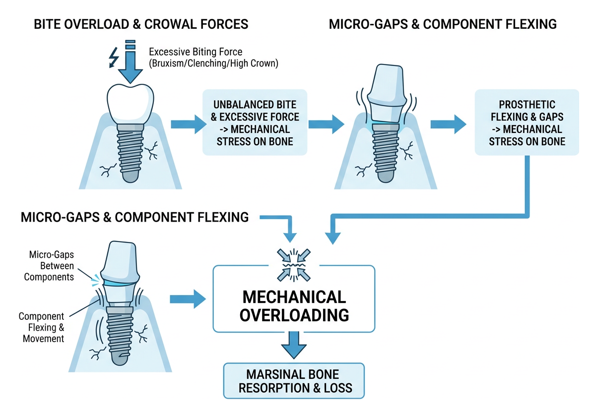
Mechanical overloading happens when an implant receives continuous, unbalanced biting forces that exceed what the surrounding bone can handle. Because dental implants lack a periodontal ligament to sense pressure, biting forces are transferred directly to the crestal bone.
Unmanaged clenching, night-time grinding (bruxism), or crowns placed slightly too high can cause micro-fractures in the surrounding bone. Additionally, surgical errors—such as overheating the bone during placement, selecting an inappropriate implant diameter, or placing components that leave micro-gaps where components flex—accelerate bone breakdown. Patients visiting our practice for second opinions often benefit from recognizing botched dental implants caused by poor prosthetic angles or misaligned fixtures placed elsewhere.
Host Factors and Systemic Issues: What Causes Bone Loss Around Dental Implants?
Systemic health conditions and immune responses play a major role in how well your body maintains bone around a titanium fixture. Research on immunological foreign-body responses shows that dental implants exist in a dynamic equilibrium with the host's immune system, and certain triggers can tip that balance toward osteoclast activation (bone breakdown).
Key systemic and host contributors include:
Uncontrolled Diabetes: Elevated blood glucose impairs microvascular circulation, weakens collagen synthesis, and increases pro-inflammatory cytokines that accelerate bone resorption.
Tobacco Use: Smoking constricts blood flow in the gums and jaw, reducing tissue oxygenation and weakening the body's defense against bacteria.
Medication Interactions: Certain medications, including long-term proton pump inhibitors (PPIs) or selective serotonin reuptake inhibitors (SSRIs), can subtly alter bone mineral metabolism.
Micro-Particle Reactivity: Microscopic titanium wear particles or surface corrosion, when combined with oral biofilm, can activate inflammatory signaling pathways (such as the RANKL pathway), causing localized bone resorption.
How Do Clinicians Diagnose and Measure Bone Loss Around Dental Implants?
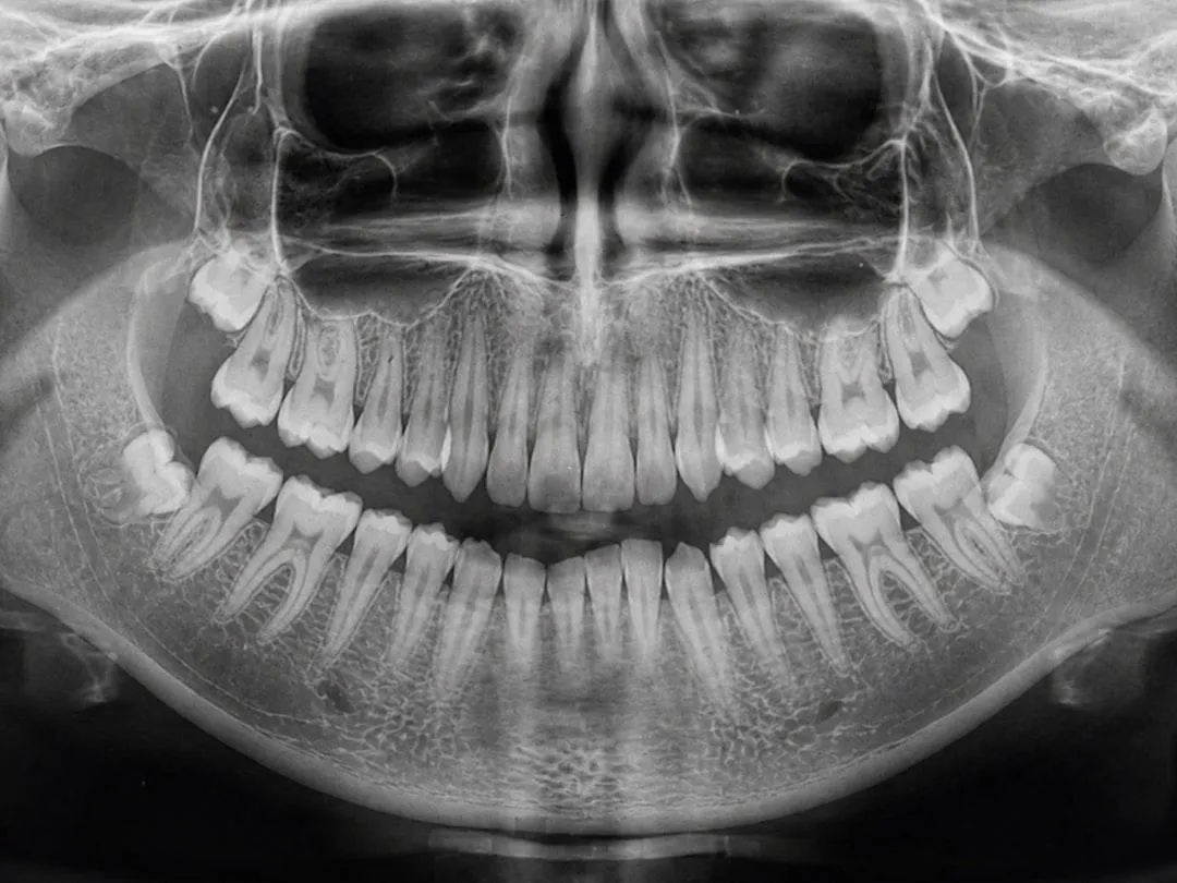
We diagnose and measure bone loss around dental implants using advanced 3D Cone-Beam Computed Tomography (CBCT) imaging, standardized digital periapical radiographs, and gentle periodontal probing. These tools allow us to evaluate bone height on all sides of the fixture with sub-millimeter precision.
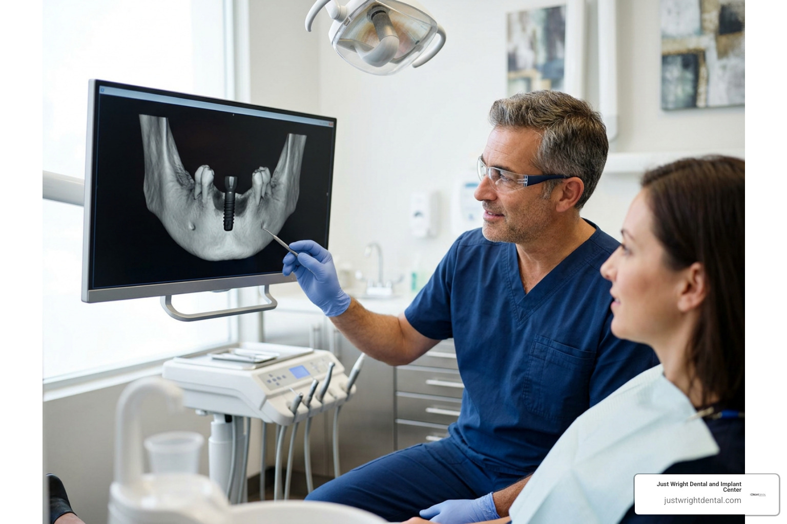
Detecting bone changes early requires precise imaging rather than guesswork. Research on measuring crestal bone loss emphasizes that relying solely on two-dimensional x-rays can miss bone loss occurring on the cheek or tongue side of an implant.
Radiographic Assessment and Baseline Calibration
Accurate diagnosis begins by comparing current digital imaging against a baseline radiograph taken when the final teeth were delivered.
3D CBCT Scans: Cone-beam 3D scans provide a 360-degree cross-sectional view of the jawbone, revealing the exact volume, density, and contour of the bone supporting each implant thread.
Parallel Periapical Radiographs: Standardized small intraoral sensors aligned parallel to the implant body ensure clear visualization of the screw threads, eliminating distortion.
Radiographic Noise Reduction: Digital calibration techniques prevent shadows or minor angle shifts from being mistaken for active bone resorption.
Clinical Symptoms and Warning Signs
While radiographic changes provide definitive measurement, clinical signs often alert patients that something is amiss:
Bleeding or Suppuration: Bleeding upon gentle probing or the presence of a small amount of pus around the gumline indicates active biological inflammation.
Gum Recession: As underlying bone recedes, the overlying gum tissue pulls back, sometimes exposing darker metal threads or abutment margins.
Implant Mobility: A healthy implant should never move; mobility is a late sign that osseointegration has been compromised.
Persistent Discomfort: Dull aching, tenderness when chewing, or a foul taste can accompany localized bone inflammation.
What Are the Most Effective Treatments for Peri-Implant Bone Loss?
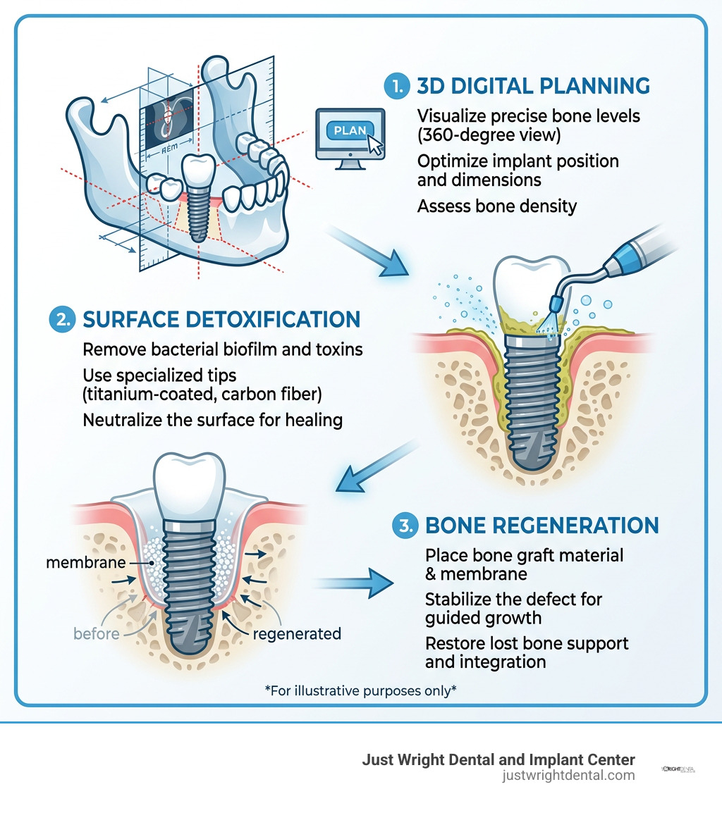
The most effective treatments for peri-implant bone loss range from non-surgical surface decontamination and localized antimicrobials for minor defects to surgical bone grafting and guided bone regeneration for advanced bone loss. The chosen approach depends on how much bone has resorbed and whether the remaining defect can hold a bone graft.
When treating bone loss, our objective is twofold: thoroughly detoxify the implant surface to eliminate bacterial toxins, and rebuild lost bone support whenever possible to preserve your dental implants.
Non-Surgical Decontamination and Maintenance
For early-stage inflammation where bone loss is minimal, non-surgical therapy is the first line of defense:
Specialized Mechanical Debridement: We use specialized ultrasonic tips (such as carbon-fiber or titanium-coated instruments) to remove biofilm without scratching the smooth collar of the implant.
Antimicrobial Irrigation: Deep irrigation with therapeutic rinses, such as chlorhexidine or targeted antiseptics, neutralizes bacteria nestled in deep pockets.
Occlusal Bite Equilibration: Adjusting the biting contacts on the crown relieves excessive physical stress, giving surrounding bone cells a stable environment to recover.
Guided Bone Regeneration and Regenerative Surgery
When bone loss has created crater-like defects around an implant, surgical regeneration is needed to rebuild structural support:
Surface Detoxification: The implant surface is cleaned and conditioned to remove microscopic bacterial endotoxins from exposed threads.
Bone Grafting Materials: We place mineralized bone graft materials, such as bioactive biphasic calcium phosphate or mineralized particulate grafts, directly into the cleaned bone defect.
Resorbable Barrier Membranes: A protective collagen membrane is placed over the graft material. This prevents fast-growing gum cells from migrating into the space, giving slower-growing bone cells time to regenerate.
Soft Tissue Grafting: Adding thick, healthy gum tissue around the neck of the restoration creates a tight biological seal that protects the underlying graft.
For extensive reconstructions, such as full-arch All On X restorations, our in-house laboratory uses 12 CAD/CAM mills and 4 3D printers to design and mill custom prosthetics with ideal contours, ensuring patients can easily clean around every support post.
How Can Patients Prevent Bone Resorption and Protect Their Implants?
Patients can prevent bone resorption around dental implants by maintaining meticulous daily plaque control, attending routine dental cleanings, wearing a custom nightguard if they clench or grind, and managing systemic health conditions like blood sugar.
Because dental implants are an investment in your health, protecting the surrounding bone requires consistent daily care and professional maintenance. You can explore further preventive strategies in our Smile Education Center - Dental Implants resource library.

Essential preventive practices include:
Targeted Interdental Cleaning: Clean around implant posts twice daily using soft interdental brushes, unwaxed implant floss, or a water flosser on a medium setting.
Custom Nightguards: If you grind or clench your teeth while sleeping, a custom-milled occlusal guard absorbs destructive forces and shields crestal bone from overload.
Tobacco Cessation: Avoiding smoking and vaping keeps blood flow healthy in the gums and supports your body's immune defenses.
Regular Professional Checkups: Schedule professional cleanings and examinations every three to six months so we can evaluate your bone levels on digital imaging and address minor pocketing before it progresses.
Frequently Asked Questions About Implant Bone Loss
Can bone loss around a dental implant be reversed?
In many cases, bone loss can be halted and partially regenerated using advanced surgical bone grafting and guided bone regeneration techniques. Success depends on catching the condition early and having a well-contained bone defect that can support graft materials. If caught in early inflammatory stages, thorough detoxification combined with soft tissue grafting can stabilize the implant.
Does bone loss mean the dental implant has failed?
Minor or localized bone loss does not automatically mean an implant has failed or must be removed. As long as the implant remains firmly integrated into the deeper jawbone without mobility, targeted decontamination and regenerative treatment can often save the fixture. An implant is generally considered failed only when bone loss is so severe that the post becomes loose or causes untreatable, recurrent infection.
How quickly does bone loss progress around an implant?
Bone loss can progress gradually over several years from mild bite stress or rapidly within months during acute bacterial peri-implantitis flare-ups. Because implants lack the natural nerve endings and periodontal ligament fibers of real teeth, bone loss often occurs without pain. This makes routine digital 3D checkups essential for catching subtle changes before significant damage occurs.
How does my overall health affect bone stability around implants?
Systemic health conditions like unmanaged diabetes, autoimmune disorders, and severe osteoporosis can impair your body's natural bone turnover and healing abilities. Maintaining tight blood sugar control, eating a nutrient-rich diet supporting bone density, and informing your dental team about any new medications help keep the bone surrounding your implants healthy.
Preserving Your Smile with Comprehensive Implant Care
Protecting the bone that anchors your dental implants is essential for enjoying a stable, confident smile for decades to come. Whether you are experiencing early gum irritation around an existing restoration, dealing with complications from treatment received elsewhere, or exploring new tooth replacement options, timely diagnosis makes all the difference.
At Just Wright Dental and Implant Center in Corsicana, Texas, we combine advanced 3D CBCT diagnostics, facially driven digital design, and in-house CAD/CAM technology to deliver precise, gentle care tailored to your anatomy. If you have concerns about bone loss or want a clear evaluation of your implant health, we invite you to schedule a Free Smile Assessment with our caring team today.
Author Bio
Dr. Justin Wright, DDS is the founder and clinical director of Just Wright Dental and Implant Center in Corsicana, Texas. With 18 years in practice and more than 10,000 implants placed, he specializes in complex full-arch reconstructions, advanced bone regeneration, and remote-anchorage implant solutions.